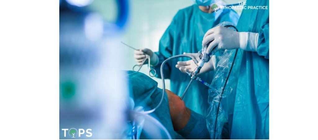
An arthroscopy is a minimally invasive surgical procedure for diagnosing and treating joint problems. Certain types of joint damage can even be repaired during arthroscopy, with miniature (pencil-thin) surgical instruments inserted through additional small cuts.
This means that you can now get a detailed look at your joint to confirm the suspected diagnosis and address any issues during the very same arthroscopy procedure. The advantage over traditional open surgery is that the joint does not have to be opened up fully.
There are two roles for arthroscopy:
- Diagnostics Arthroscopy: Allows evaluation of the joint through real-time assessment of the joint
- Therapeutics Arthroscopy: Allows minimally invasive surgery to be performed to treat joint disease
Arthroscopy can be performed on the hip, knee, ankle, shoulder, elbow and wrist joints.

Common Conditions Where an Arthroscopy is required
Knee
Shoulder
- Rotator cuff tear
- Removal or repair of the labrum
- Repair of ligaments
- Removal of inflamed tissue or loose cartilage
- Repair for recurrent shoulder dislocation
How do I Prepare
Exact preparations depend on which joint(s) your surgeon is examining or repairing. In general, you should:
- Avoid certain medications based on your doctor’s advice
- Fast beforehand, up to eight hours before your procedure
- Always take guidance from your doctor on the actual preparation he recommends
During the Arthroscopy Procedure
Types of anesthesia: Varies by procedure
a. Spinal anesthesia. This is the most common form of anesthesia. The bottom half of your body will be numbed and you remain awake during the entire procedure.
b. General anesthesia. Depending on the length of the operation, it may be necessary for you to be asleep during the procedure. General anesthesia is administered through a combination of intravenous medications and inhaled gases.
2. Steps involved
- One small cut (incision) is made for insertion of the viewing device – a narrow camera attached to a fiber-optic video camera.
- Your doctor will fill the joint with a sterile fluid to improve the view of the joint and to control bleeding.
- The view captured by the camera will be transmitted and projected onto a high-definition video monitor.
- Additional small cuts at around the joint will allow the doctor to insert miniature surgical tools to grasp, cut, grind and provide suction as needed for the procedure.
- Incisions will be small enough to be closed with one or two stitches, or with narrow strips of sterile adhesive tape.
3. Time taken
Arthroscopic surgery usually does not take long. You will then be taken to a separate room to recover for a few hours before going home.
After Arthroscopy
1. Medications.
Your doctor may prescribe medication to relieve pain and inflammation.
2. R.I.C.E. – Rest, Ice, Compress and Elevate
You may find it helpful to rest, ice, compress and elevate the joint for several days to reduce swelling and pain.
3. Protection.
- You might need to use walking aid temporarily — e.g. crutches for comfort and protection.
- You might also need to use splints temporarily – e.g. knee brace for comfort and protection
4. Exercises.
You might be prescribed physical and rehabilitation therapy to help strengthen your muscles and improve the function of your joint.
5. Checks to be done by staff
- Monitoring your blood pressure, breathing, and pulse
- Checking of nerves and pulses near the operated area

